Johann Kruger MD M Med(Oph) FCS SA (Oph) FRCS Ed(Oph ) FWCRS Tygervalley Eye & Laser Centre, Cape Town, South Africa.
Planning
- Manifest refraction by optometrist done
- Selecting patients with advanced Keratoconus with < 20/50 vision
- Patients correcting to 20/20 are not operated
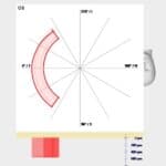
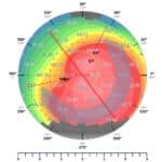
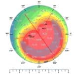
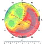
MS 39 Sagittal map is used Anterior segment OCT Customisation:
- Single rings

- Double rings
- Tissue ring designed to cover and flatten the steep areas
- Edges are tapered
- An asymmetric ring is cut with the laser

Methods
Selecting patients with advanced Keratoconus with < 20/50 vision Patients correcting to 20/20 are not operated Cornea – Corneal tissue is sourced from The Bay Tissue bank in SA – Or from The Lions Eye bank in the USA 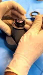 Cornea preparation:
- Cut with a low energy Femtosecond laser
- Anterior chamber maintainer is used –
- Epithelium is removed with a sponge
- The cornea is preserved in Optosol solution
- The cornea is removed from the fridge to bring to room temperature
- Bowman’s is methyline blue dyed with a marker pen
- The segment sizes are then cut and placed on a degree marker with Bowman’s ring inwards
- Constant thickness cut but varying the width of the segment
- The patient ring tunnels are cut later with the femtosecond laser once segments dry
Methods
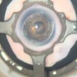
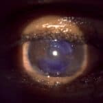
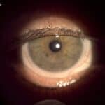
- Principle is to cover the steepest area with the segment.
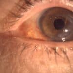
- The patient’s cornea is marked with a marking pen
- Digital marking system used to mark where tissue inlay must be placed on the patient.
- The patient ring tunnels are cut then with the femtosecond laser, once the segments are dry and ready for insertion.
Plans
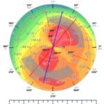
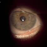
Case One
- 44 y.o lady with bilateral advanced Keratoconus
- No previous ophthalmic surgical history
- OD: UCVA 20/100, BCVA 20/40 (-1.50/ -3.00 x 7)
- OS: UCVA FC at 3m, BCVA 20/100 (-8.00/ -8.00 x 173)
- Bilateral CAIRS was performed in June 2025 – single segment inserted OU
OD:1200um x 600um
- Tunnel width: 1.50mm
- Tunnel depth: 250um
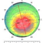
OS:1200um x 600um
- Tunnel width: 1.50mm
- Tunnel depth: 250um
Outcome OD: UCVA 20/25 1 week post-op (0.00/ -1.75 x 174)
Outcome OS: UCVA 20/60 1 week post-op (-4.75/ -3.00 x 12)
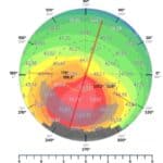
Case Two
- 23 y.o lady known newly diagnosed with Keratoconus
- No previous ophthalmic surgical history
- OD: UCVA 20/200, BCVA 20/25 (-0.75/ -1.25 x 75). X
- OS: UCVA 20/125, BCVA 20/80 (-4.00/ -1.75 x 25)
- Left CAIRS was performed in June 2025 –
- Single segment inserted – 1200um x 600um
- Tunnel width: 1.50mm
- Tunnel depth: 300um
- Outcome: UCVA 20/25 2 weeks post-op

Case Three
- 40 y.o gentleman known with Keratoconus
- Bilateral CXL x3 previously
- OD: UCVA FC at 1m, BCVA 20/32 (-11.50/ -4.00 x 65)
- OS: UCVA FC at 2m, BCVA 20/50 (-9.50/ -6.00 x 115)
- Left CAIRS was performed in June 2025
- single segment inserted – 1200um x 600um
- Tunnel width: 1.50mm
- Tunnel depth: 250um
- Outcome: UCVA 20/320 2 weeks post-op, BCVA 20/40 (-2.00/ -3.25 x 120)
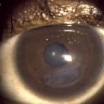
Case Four
- Poor vision, not able to get useful vision with correction
- Had CME previously OU requiring Avastin IVT injections
- OD: UCVA 20/25, BCVA 20/20 (-0.75/ -0.75 x 75)
- OS: UCVA 20/200, BCVA 20/63 (-1.25/ -4.25 x 120)
- 64 y.o lady known with Keratoconus and previous corneal graft with Ectasia temporally in the graft
- Customised Left CAIRS was performed in June 2025
- single segment inserted with tapering width
- Segment width: 1.3mm tapering to 1.0mm
- Segment thickness: 550um
- Tunnel width: 1.60mm
- Tunnel depth: 250um
- Outcome: UCVA 20/50 2 weeks post-op, BCVA 20/40 (+0.25/ -6.00 x 165)
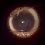
Case Five
- Left CAIRS was performed in June 2025
- 2 segments inserted – 1200um x 600um
- Tunnel width: 1.40mm
- Tunnel depth: 250um
- Outcome: UCVA 20/32 2 weeks post-op (+0.50/-1.50 x 175)